Philadelphia / Bucks County LASIK Surgeon – Dr. James S. Lewis
Eye News Volume II Issue 11

Volume II Issue 11
Originally published, Sunday, April 29, 2001
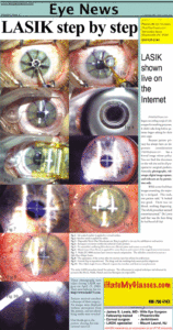
LASIK step by step

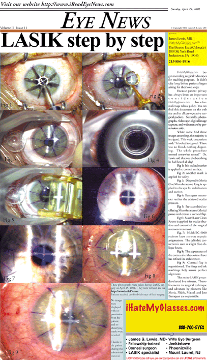
Fig 1: Ink soaked marker is applied to corneal surface.

Fig 2: Another mark is applied for safety.

Fig 3: Disposable Moria One Microkeratome Ring is applied to the eye for stabilization and suction.

Fig 4: Barraquer tonometer verifies the achieved ocular pressure.

Fig 5: Pre-assembled oscillating Microkeratome (Moria) passes and creates a corneal flap.

Fig 6: Mastel Laser Clean Room is applied for ocular fixation and control of the surgical microenvironment.

Fig. 7: Nidek EC-5000 excimer laser corrects myopic astigmatism. The cylindric correction is seen as a light blue oblique beam.
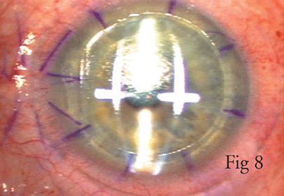
Fig 8: The appearance of the cornea after the excimer laser has refined its architecture.
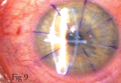
Fig 9: Corneal flap is respositioned. The hinge and ink markings help assure perfect alignment.
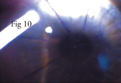
Fig 10: Fiber Optic Light Source (Mastel) inspects the interface and final corneal placement.
The entire LASIK procedure lasted five minutes. The refinements in surgical technique and advances by pioneers like Moria, Nidek, Mastel, and Jose Barraquer are responsible.
These photographs were taken during LASIK surgery on April 25, 2001. They were webcast live via
http://www.lasikTV.com.
http://www.lasikTV.com.
Patients received unedited videotape of their surgery.
No images were displayed without permission from the patient, and no identifying marks were revealed.
Our thanks go to the patient sharing this educational material.
LASIK shown live on the Internet
iHateMyGlases.com began recording surgical videotapes for teaching purposes. It didn’t take long before patients began asking for their own copy.
Because patient privacy has always been an important consideration iHateMyglasses.com has a formal image release policy. You can find this document on the web site and in all pre-operative surgical packets. Naturally, photographs, videotape, digital image capture, and webcasts are by permission only.
While some find these images unsettling, the majority is intrigued. This week, one patient said, “It looked too good. There was no blood, nothing disgusting. The whole procedure seemed somewhat unreal.” Dr. Lewis said that was the best thing he had heard all day!
As a highly regarded Philadelphia Epi-LASIK and LASIK surgeon, Dr. Lewis strives to deliver the best possible vision correction results for each one of his patients. In addition to performing LASIK eye surgery in Philadelphia, Dr. Lewis has many years’ experience treating Philadelphia ICLs patients. In fact, he was one of the first surgeons in Pennsylvania to perform implantation of these sophisticated collamer lenses.

