Philadelphia / Bucks County LASIK Surgeon – Dr. James S. Lewis
Eye News Volume II Issue 7

Volume II Issue 1
Originally published, Sunday, April 1, 2001
LASIK after Dark: How to reduce glare
Glare is an important consideration for LASIK patients and LASIK surgeons. Early complaints of halos or difficulty with night driving have inspired refinements in laser technique. Measurement of pupil diameter in a dark room is now part of the standard LASIK evaluation.

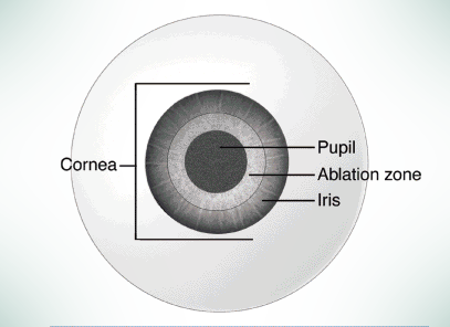
An ideal ablation zone is larger than the diameter of the pupil. This reduces the chance of glare and halos.
Producing an ablative zone (lasered area) larger than the pupil diameter should reduce these symptoms. All light rays enter the eye focused, so no halos are produced. Initially, excimer lasers could only produce ablative zones between 4.5 and 6.0 millimeters. Because most pupils dilate from 5 to 9 millimeters in darkness, it is not surprising early laser vision correction patients experienced glare.
Virtually all popular excimer laser manufacturers have upgraded their systems. Patients with large pupils still represent a challenge to the LASIK surgeon. “You can’t simply continue to increase the size of the ablative zone. You will sacrifice too much corneal tissue and risk ectasia [weakening and bulging of the cornea],” explained Dr. Lewis.
Jose Barraquer, MD, of Bogota Columbia, the grandfather of LASIK suggested that 250 microns of untreated cornea remain after surgery. Ultrasonic pachymetry measures each patient’s corneal thickness prior to surgery. Measurements range from 420 microns to 600 microns.
“We routinely calculate the residual cornea before any LASIK procedure. Enhancements are no exception. We must temper our desire to minimize glare with our need to leave the patient with a strong and healthy cornea,” Lewis continued.
LASIK surgeons today divide the ablative zone into an inner optical zone and a surrounding transition zone. The optical zone corrects the patient’s vision. The transition zone removes less tissue and smooths the area between the optical zone and the untouched peripheral cornea.
Lewis claims, “One of the truly wonderful advantages of our Nidek laser is its ability to tailor the optical zone and the transition zone for each patient. I can consider pupil size, refractive error, and corneal thickness and select the ideal ablation pattern.”
Another advantage of separate optic and transition zones is the reduction of regression. The corneal contour is gently curved. The body tolerates this shape and exhibits little or no tissue response. Enhancement rates are significantly reduced.
Laser manufacturers have helped by improving their alignment and aiming systems. The attached surgical microscope helps the doctor position the laser.
“The fiber optic light source and the fixation capabilities of our system make me confident. I know the laser energy will go exactly where it should,” commented Lewis.
Medicine and industry continue to improve LASIK. 20/20 acuity is no longer the gold standard. Patients want excellent vision in all lighting conditions.
Happy Faces

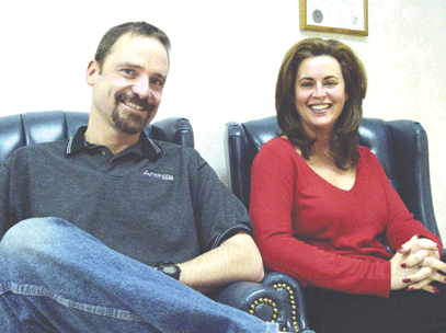
Karen and David Thompson relax for a photo during one of their post-operative LASIK visits.

Over the years, Dr. Lewis has performed numerous LASIK procedures that have helped his Philadelphia and Bucks County laser eye surgery patients eliminate or significantly reduce their dependence on glasses. In addition to LASIK, Dr. Lewis performs Epi-LASIK and is renowned as one of the most experienced Philadelphia / Bucks County Visian ICL experts.

